Diagnosing Glaucoma
The only sure way to diagnose glaucoma is with a complete eye exam. A glaucoma screening that only checks eye pressure is not enough to find glaucoma.
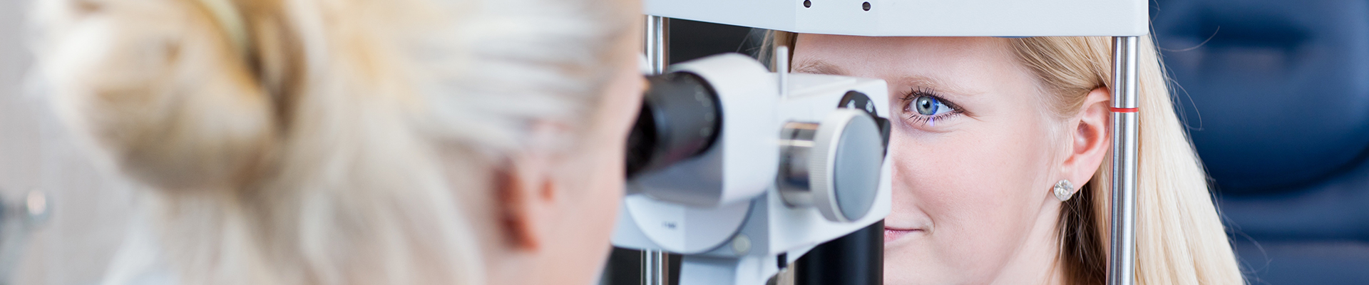
During a glaucoma exam, your ophthalmologist will perform:
- Applanation Tonometry – A measurement of your eye pressure.
- Gonioscopy – An inspection of the drainage area of your eye to determine if it is open, narrow or closed. A special mirrored lens is used to view this structure.
- Ophthalmoscopy – A microscopic examination technique used to evaluate the optic nerves to look for signs of damage from glaucoma. This is best performed after pupil dilation.
- Visual Field Testing – A test of your peripheral (side) vision. This allows detection of subtle changes to the vision of which you may not be aware and allows for monitoring of changes over time.
- Pachymetry – A measurement of your corneal thickness. This test allows further interpretation of your intraocular pressure measurement.
- Computerized Optic Nerve Imaging – There are several different devices currently available to assess the optic nerves.
These include:
- Stereoscopic Photographs of the Optic Nerves: This provides three-dimensional photographs of the surface of your optic nerves to look for signs of damage from glaucoma.
- Optical Coherence Tomography (OCT): This provides computer-generated measurements of the retinal nerve fibers that join together to form the optic nerve. Damage to these fibers is an extremely sensitive marker for glaucoma. This test allows a very accurate comparison of your retinal nerve fibers from year to year.
- Heidelberg Retina Tomography (HRT): This provides a computer-generated three-dimensional map of the surface of the optic nerve to look for signs of damage from glaucoma. This test allows a very accurate comparison of your optic nerve from year to year.